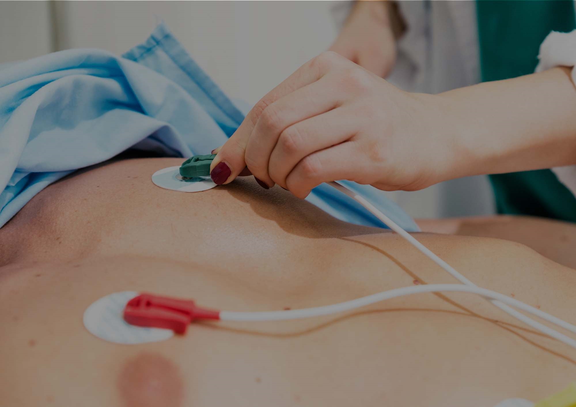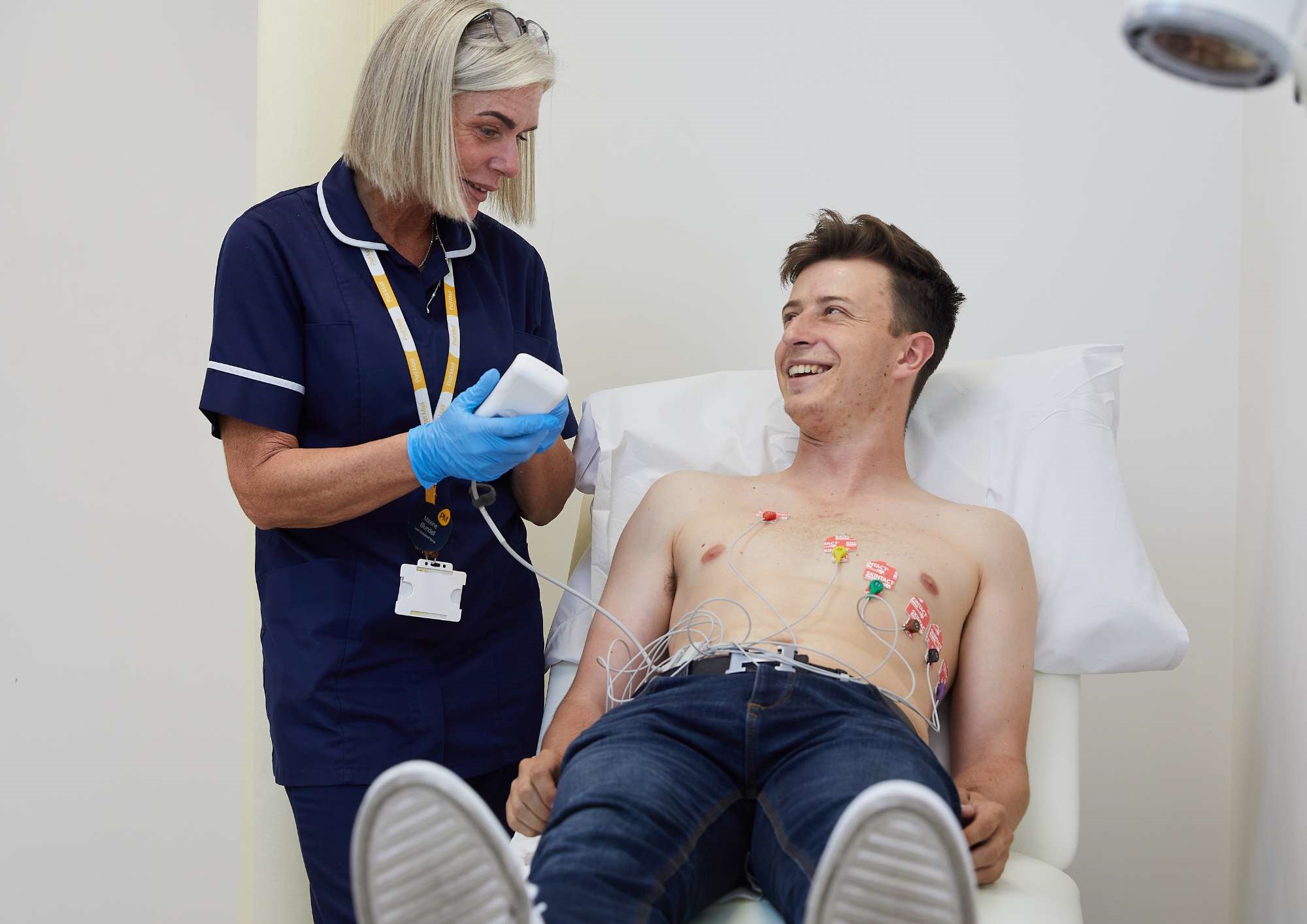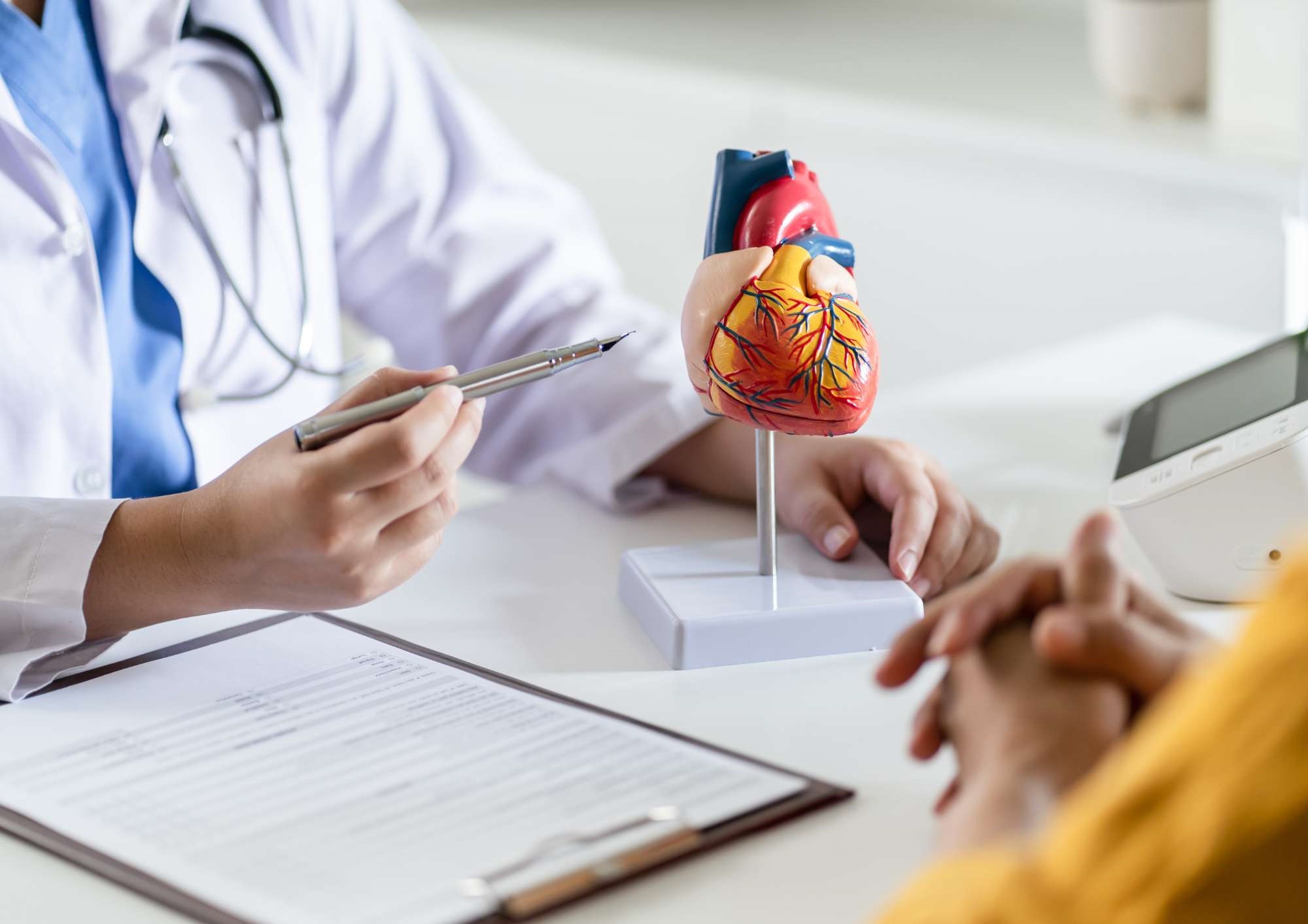
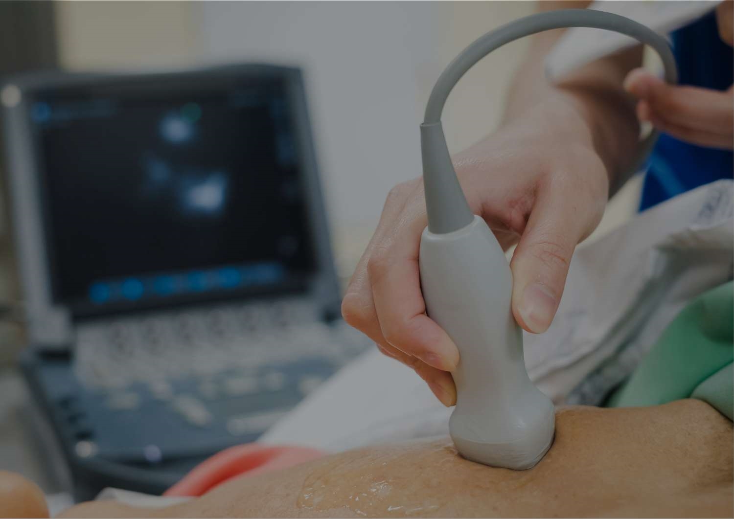
An ECHO, also commonly known as an echocardiogram, is a non-invasive and highly effective imaging technique used to produce detailed pictures of the heart using sound waves. It is widely used in diagnosing and managing a variety of heart conditions and provides invaluable insight into heart function, structure, and overall health. Safe, quick, and painless, an ECHO is an essential tool in the early detection and ongoing management of heart disease, offering a clear picture of the heart’s function and helping doctors make informed decisions regarding treatment and care.
What are the benefits of an ECHO
Early detection of heart conditions
An ECHO is one of the most effective tools for the early detection of heart conditions, some of which may not exhibit symptoms initially. By assessing the heart's structure and function, an ECHO can identify abnormalities such as heart valve disease, congenital heart defects, and a weakened heart muscle. Early detection of such conditions can significantly improve the prognosis and allow for prompt treatment, which is often less invasive and more effective when started early.
Monitoring heart function
An ECHO allows doctors to evaluate the efficiency of the heart's pumping action. By measuring the ejection fraction, the percentage of blood the heart pumps out with each beat, it helps to assess the heart’s ability to circulate blood throughout the body. This is crucial for patients with heart failure, as it can help determine the severity of the condition and inform treatment decisions, such as adjustments to medication or recommendations for additional therapies.
Assessing heart valve health
Heart valve disease is a common issue that can lead to serious complications if left untreated. An ECHO provides a clear view of the heart valves, allowing doctors to assess whether they are functioning properly or if there is any regurgitation (backflow) or stenosis (narrowing). Identifying issues with heart valves early on helps doctors determine the need for medical intervention, such as valve repair or replacement, to prevent complications like stroke, heart failure, or infection.
Detecting blood clots and fluid accumulation
An ECHO can identify the presence of blood clots within the heart, particularly in the atria, which may lead to life-threatening conditions such as stroke or pulmonary embolism. Additionally, it can help detect fluid accumulation around the heart, a condition known as pericardial effusion, which may indicate inflammation or infection. Early detection through an ECHO enables timely intervention, reducing the risk of further complications.
No radiation exposure
Unlike other imaging techniques such as CT scans or X-rays, an ECHO uses sound waves to create images, making it a completely safe, non-invasive procedure that does not expose patients to radiation. This makes it a highly recommended choice for pregnant women or individuals who may need frequent heart monitoring over time, as it can be safely performed without long-term health risks.
Detecting structural heart issues
An ECHO is also vital in identifying any structural abnormalities in the heart, such as an enlarged heart (cardiomegaly), thickened heart walls, or congenital defects. These issues may not show obvious symptoms in their early stages but can progress into more severe heart problems if not diagnosed and treated promptly. An ECHO allows for comprehensive imaging of the heart, offering insight into how well the heart is functioning and whether any abnormalities need to be addressed.